First direct observation of how cells react to a magnetic field
Scientists in Japan have for the first time observed how living cells appear Magnetic fields react. Your research could prove crucial to understanding how animals, from birds to butterflies, use the Earth's magnetic field to navigate. It may also be possible to find out whether weak electromagnetic fields can affect our health.
Many animal species have the ability to Magnetoreception, so to perceive the earth's magnetic field. They use them to navigate the planet, especially long-distance hikes. However, the mechanisms behind the magnetic "sixth sense" are poorly understood. Japanese scientists from the University of Tokyo have taken a step towards a better understanding of magnetic reception. In their laboratory, they observed how living, non-genetically modified cells react to magnetic fields. The results were in the journal Proceedings of the National Academy of Sciences released. The researchers' work can help us understand how animals use magnetic fields for navigation and whether such fields can affect human health.
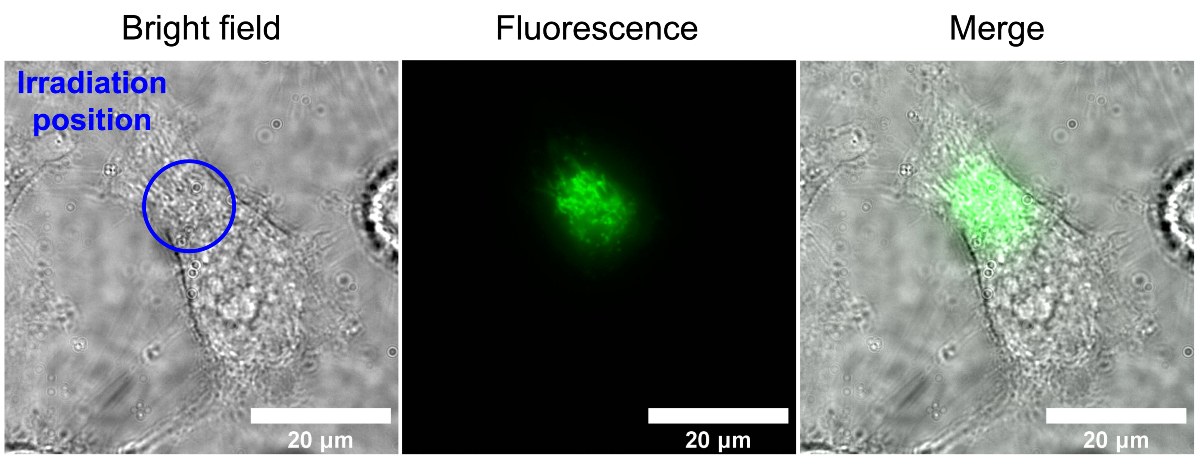
Image source: www.u-tokyo.ac.jp/content/400152121.jpg
Magnetoreception in living cells
Scientists have long suspected that the Earth's magnetic field could affect animal behavior. They were stimulated by the simple observation that a magnet can attract or repel electrons. This in turn allows the conclusion that magnetic fields can influence chemical reactions in cells.
When certain molecules are excited with light, an electron can jump from one to the other and form two molecules with single electrons, a so-called Radical pair. The individual electrons can exist in one of two states, which differ in spin. When the radicals have the same spin, their subsequent chemical reactions are slow, while pairs of radicals with opposite spins can react faster. Magnetic fields can influence the spin of electrons and thus directly influence chemical reactions with radical pairs.
In recent years, scientists have identified several proteins that Cryptochromes to be named. These are blue light sensitive photoreceptors found in both plants and animals. They are also sensitive to magnetic fields.
In previous experiments, scientists observed that genetic manipulation of Cryptochromes in fruit flies and cockroaches the magnetic "sixth senseOther studies have shown that geomagnetic navigation in birds and other animals is induced by light that is necessary for the formation of the radicals mentioned above. But no one has measured the chemical reactions within a living cell that are directly involved change due to a magnetic field.
Cell autofluorescence
Woodward and his colleagues worked with HeLa cells, a cervical cancer cell-derived cell line commonly used in research laboratories. The scientists were particularly interested in the ones present in them Cryptochrome subunits, called flavin, which naturally fluoresce when exposed to blue light.
Flavins are normally used by cells to detect light, but they also provided a fantastic way for scientists to do that Magnetoreception to investigate. That's because different conditions can affect the amount of light they emit, including magnetic fields. When light falls on a flavin, the flavin emits its own light or produces radical pairs. The fluorescence depends on how quickly the radical pairs react.
The team from the University of Tokyo hoped to observe biological magnetoreception by observing the autofluorescence of cells when an artificial magnetic field was added to their environment.
According to the study authors, autofluorescence is common in cells. To the Flavin autofluorescence To isolate, the researchers used lasers to illuminate cells with light of a specific wavelength, and then measured the wavelengths of the light emitted by the cells to ensure that it matched the characteristic values of flavin autofluorescence.
Try
The cells were irradiated with blue light for about 40 seconds. The researchers irradiated the cells with a magnetic field every four seconds and measured the changes in fluorescence intensity. Analysis of the visual data from the experiments showed that the fluorescence of the cells decreased by about 3,5 percent each time the magnetic field passed through the cells.