Innovative ultrasound patch allows 48-hour monitoring of the body
 The Ultrasound examination (USG) is a safe, non-invasive method that allows a physician to obtain valuable information about a patient's internal organs. However, it currently requires the use of large, bulky and expensive equipment that is only available in doctor's offices. Engineers at MIT have created a miniature ultrasound systemdeveloped that can be stuck to the skin like a band-aid and for 48 hours provides images.
Tests on volunteers have shown that the device adheres well to the skin and provides good quality images of large blood vessels and deeper organs. In addition, it was possible to change the organs recorded during various activities when subjects were sitting, standing, walking, or cycling.
At this stage, the prototype needs a wired connection to a device that converts the reflected waves into images. However, as the developers assure us, it could be useful even now. For example, it could be used in a hospital to continuously monitor a patient without a doctor having to perform the examination.
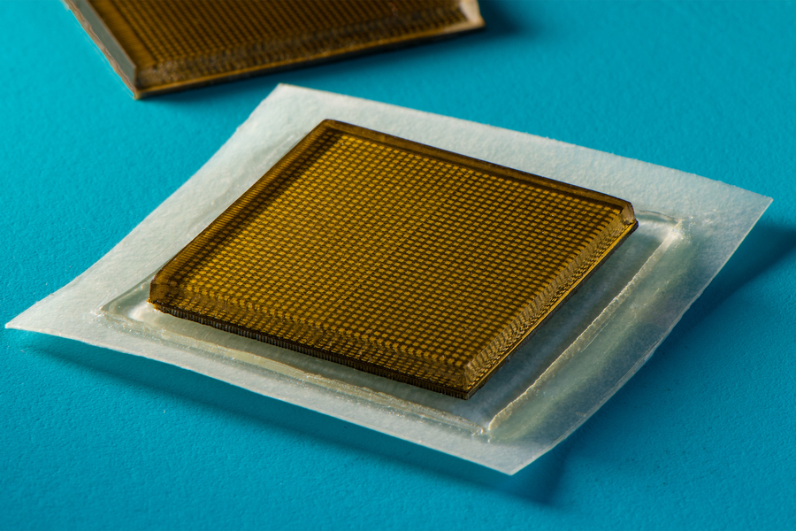
Image source: MIT ; Which
Work is currently underway on the Patches equipped with wireless connections. Such ultrasound patches could then be applied in the doctor's office or even at home and send data to, for example, the patient's phone, from where the image can be further transmitted or in Real-time analysis by artificial intelligence algorithms could. We believe we have ushered in a new era in mobile imaging. With just a few band-aids, it will be possible to observe a person's internal organs, says the study's lead author, MIT engineering professor Xuanhe Zhao.
Until now, it has not been possible to develop a device that has a high quality Long-term ultrasound monitoring allows. A handheld ultrasound imaging device would have tremendous potential for the Diagnostics. However, current ultrasound patches only provide relatively low-resolution images and cannot image deep-lying organs, says MIT's Chonghe Wang.
The researchers from Massachusetts have succeeded in combining a flexible adhesive layer with a rigid transducer arrangement. This allowed the device to be placed on the skin while maintaining the relative position of the transducers for better imaging. In previously used solutions of a similar nature are the transducer arrays flexible, which changes the position of the transducers relative to each other, resulting in image deformation and lower resolution.
The self-adhesive item the device consists of two elastomer layers, between which an elastic, stretchable gel is encapsulated. The elastomer protects the gel from drying out, explains Xiaoyu Chen, co-author of the device. The lower elastomer layer is self-adhesive, the upper layer houses the sensors. The entire device has a surface area of approximately 2 cm2 and a thickness of 3 mm.
When tested on volunteers, the wearable ultrasound device adhered well to the skin for 48 hours and provided good images while the test subjects sat, stood, ran on a mechanical treadmill, rode a stationary bike, and lifted weights. For example, the researchers were able to determine the difference in blood vessel diameter observed between a sitting and a standing subject. The disks recorded changes in the shape of the heart with exercise or changes in the stomach with drinks. And when participants lifted weights, the ultrasound detected bright marks in the muscles that indicated temporary microtrauma. For example, we can recognize the moment when an exercise is overloading the muscles and stop it, Chen explains.
The researchers now aim to develop a wireless version of the ultrasonic patch to be developed that can be used in the doctor's office or at home, not only to monitor organs, but also to monitor, for example, the course of cancerous tumors or the development of a fetus.